Images 05. Muscular System Basic Human Anatomy
The Muscular System Learn about the three types of muscle as you use our 3D models to explore the anatomical structure and physiology of human muscles. And don't worry, we'll explain the naming of skeletal muscles, too. By: Tim Taylor Last Updated: Oct 10, 2021 2D Interactive NEW 3D Rotate and Zoom Anatomy Explorer HEAD AND NECK
Muscular System Anatomy and Physiology Nurseslabs
Muscular system anatomy and physiology Sliding filament model of muscle contraction Muscle contraction Neuromuscular junction and motor unit Osmosis Muscles high-yield notes offers clear overviews with striking illustrations, tables, and diagrams. Make learning more manageable.
The Musculoskeletal System (Structure and Function) (Nursing) Part 4
Muscular System Anatomy, Diagram & Function | Healthline human body maps muscular system Muscular The primary job of muscles is to move the bones of the skeleton, but muscles also.
Muscles Diagrams Diagram of muscles and anatomy charts HubPages
The coracobrachialis is the smallest of the three muscles that attach to the coracoid process of the scapula. (The other two muscles that attach here are the pectoralis minor and the short head of the biceps brachii.) It is situated at the upper and medial part of the arm. It is supplied by the musculocutaneous nerve.
Muscular System, Front Hilmers Studios
Each skeletal muscle has three layers of connective tissue (called "mysia") that enclose it and provide structure to the muscle as a whole, and also compartmentalize the muscle fibers within the muscle ( Figure 10.3 ).

the muscular system (lesson 0386) TQA explorer
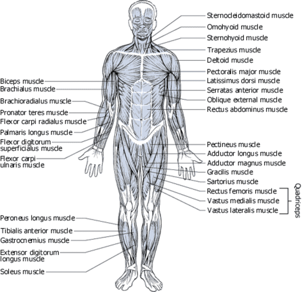
Discover the muscle anatomy of every muscle group in the human body. Find the best weight lifting exercises that target each muscle or groups of muscles. You can click the links in the image, or the links below the image to find out more information on any muscle group. Abdominals | Biceps | Calves | Chest | Forearm | Glutes | Hamstrings

Clip Art Muscular Diagram Muscular System Diagram Major Muscles, HD Png Download , Transparent
Leg Muscles Anatomy, Function & Diagram | Body Maps Plan Human body Muscular System Muscles Muscles The majority of muscles in the leg are considered long muscles, in that they.

Labeled Muscle Diagram Chart Free Download
Muscular System / In these topics. Muscles. Brought to you by Merck & Co, Inc., Rahway, NJ, USA (known as MSD outside the US and Canada)—dedicated to using leading-edge science to save and improve lives around the world. Learn more about the MSD Manuals and our commitment to Global Medical Knowledge.
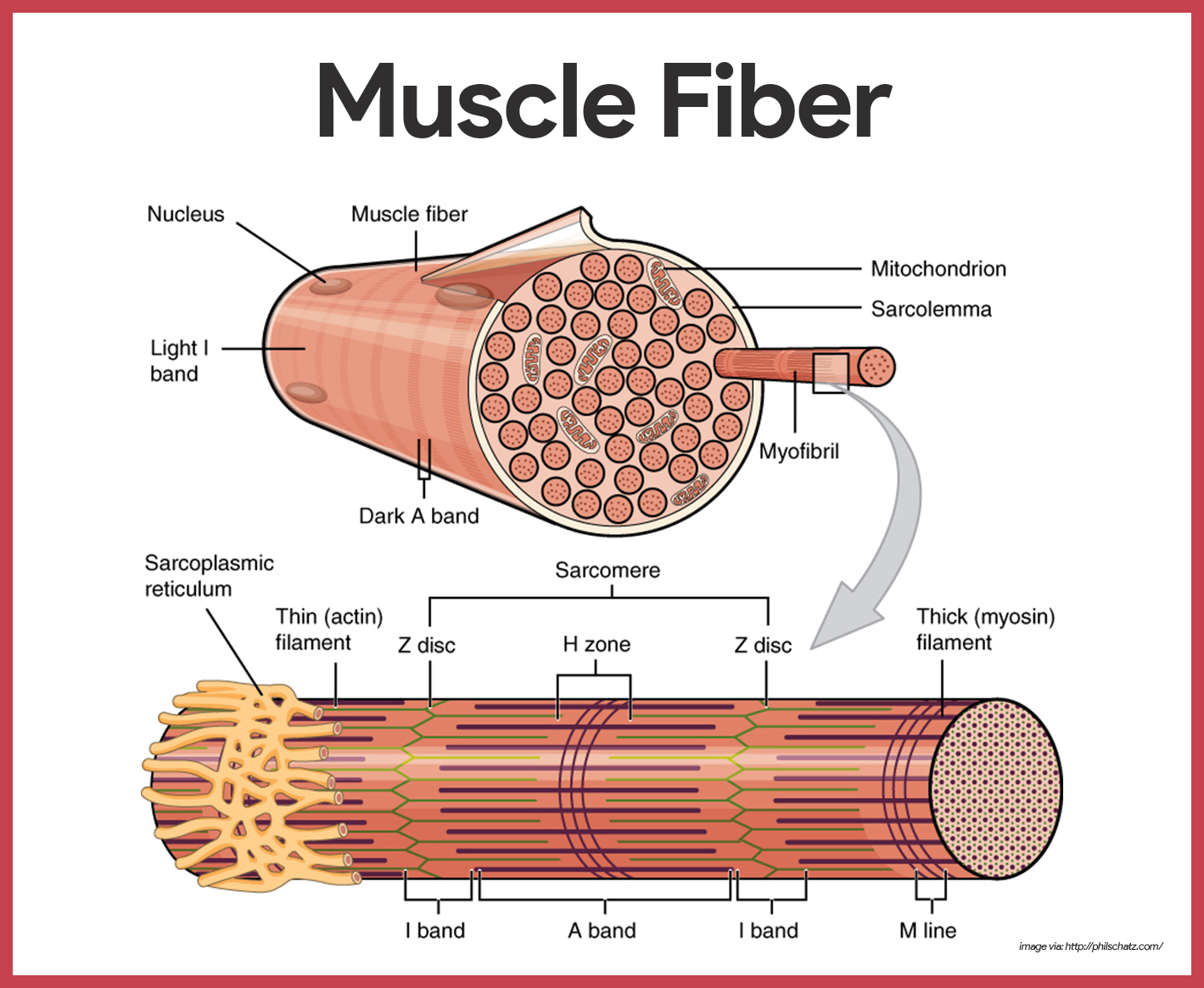
What Is the Microscopic Anatomical Makeup of Skeletal Muscles
Skeletal Muscle Fibers Because skeletal muscle cells are long and cylindrical, they are commonly referred to as muscle fibers (or myofibers). Skeletal muscle fibers can be quite large compared to other cells, with diameters up to 100 μ m and lengths up to 30 cm (11.8 in) in the Sartorius of the upper leg.

Pin on Anatomy Diagrams
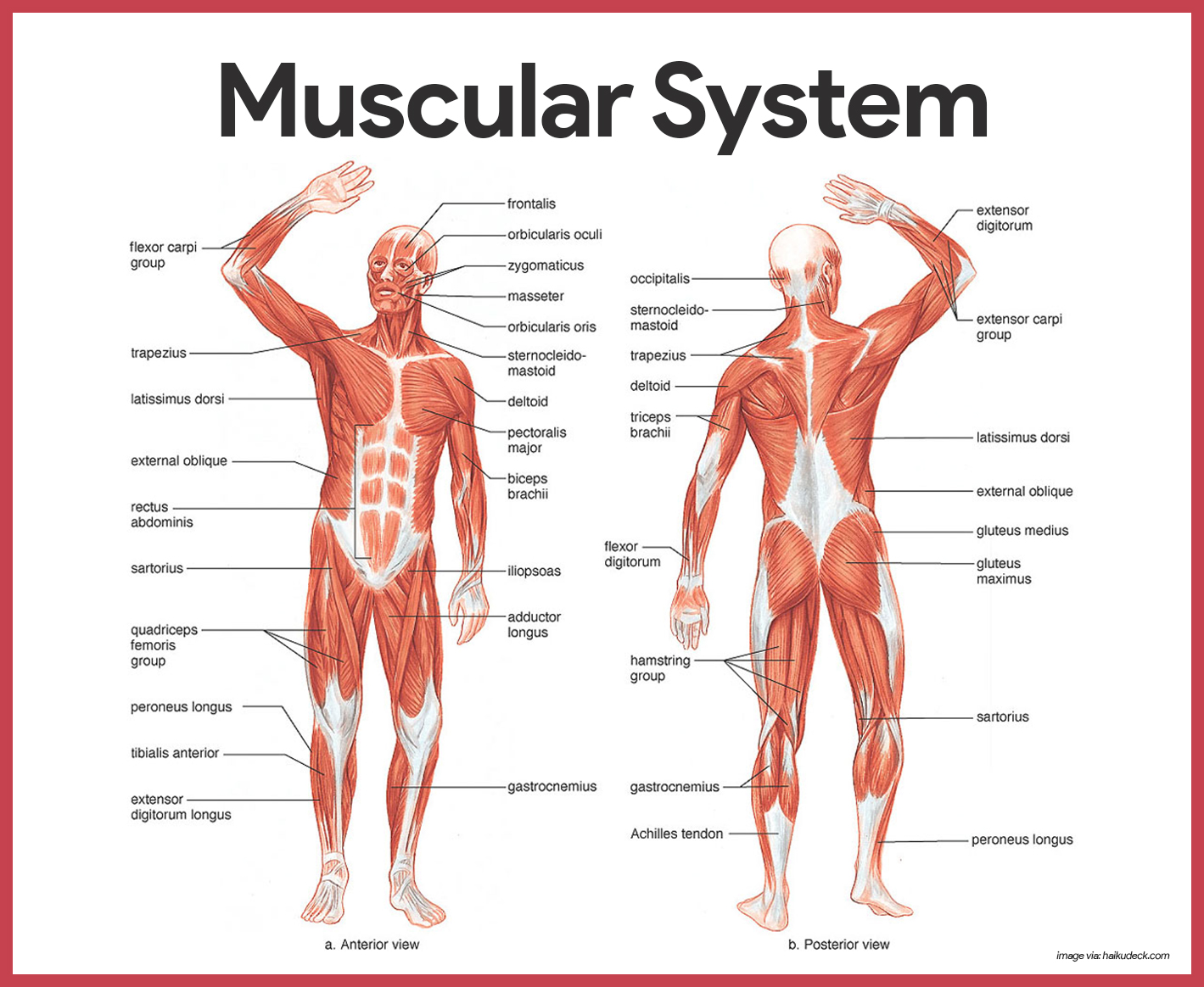
Now that we know about muscle tissue, let's see how this is arranged to form the muscular system, the incredible network of muscles that covers the skeletal.

Muscle Diagram Most Important Muscles Of An Athletic Male Body Anterior And Posterior View
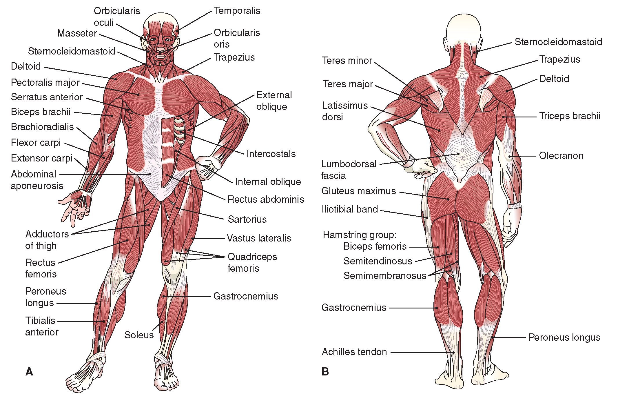
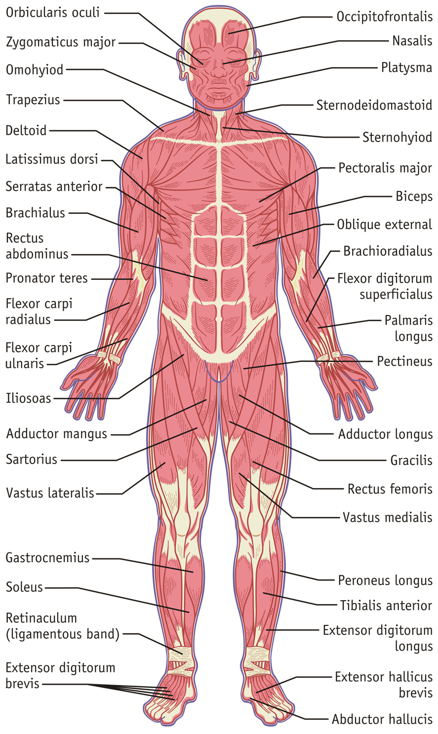
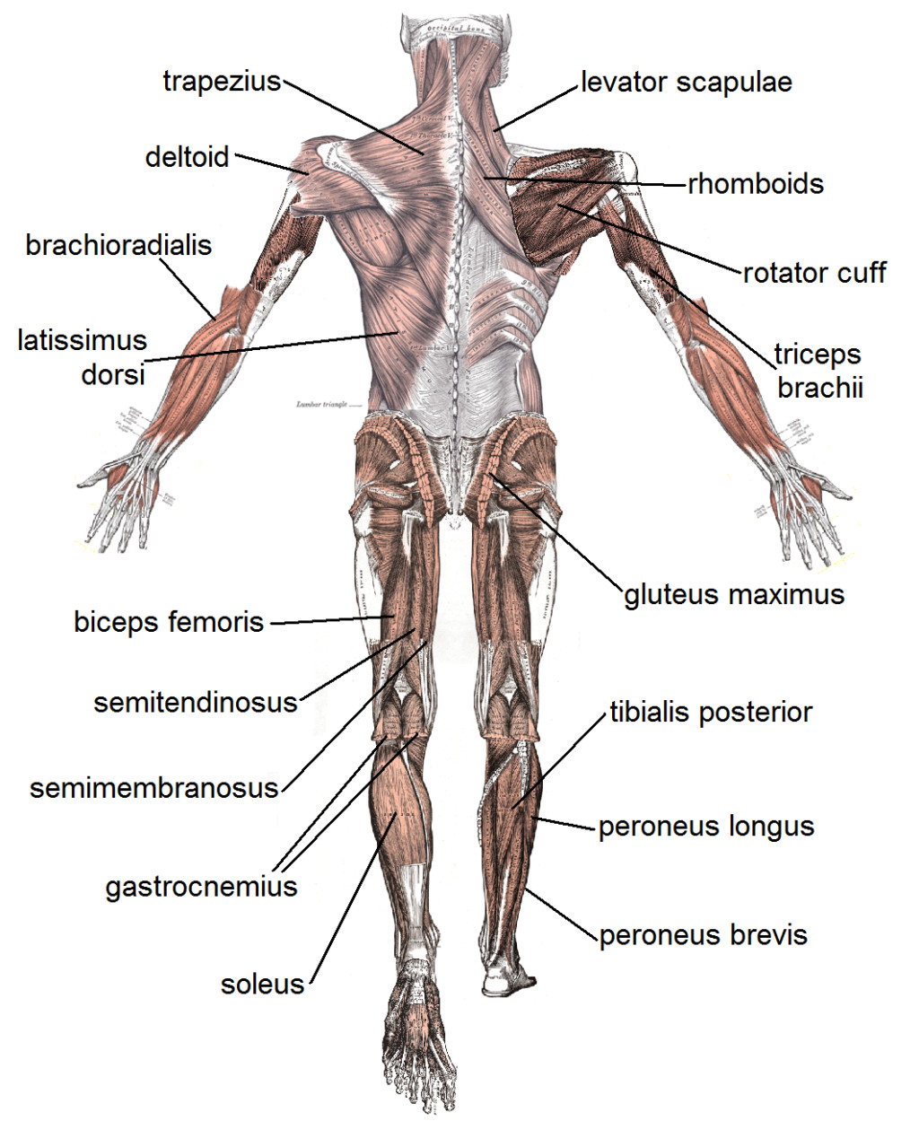
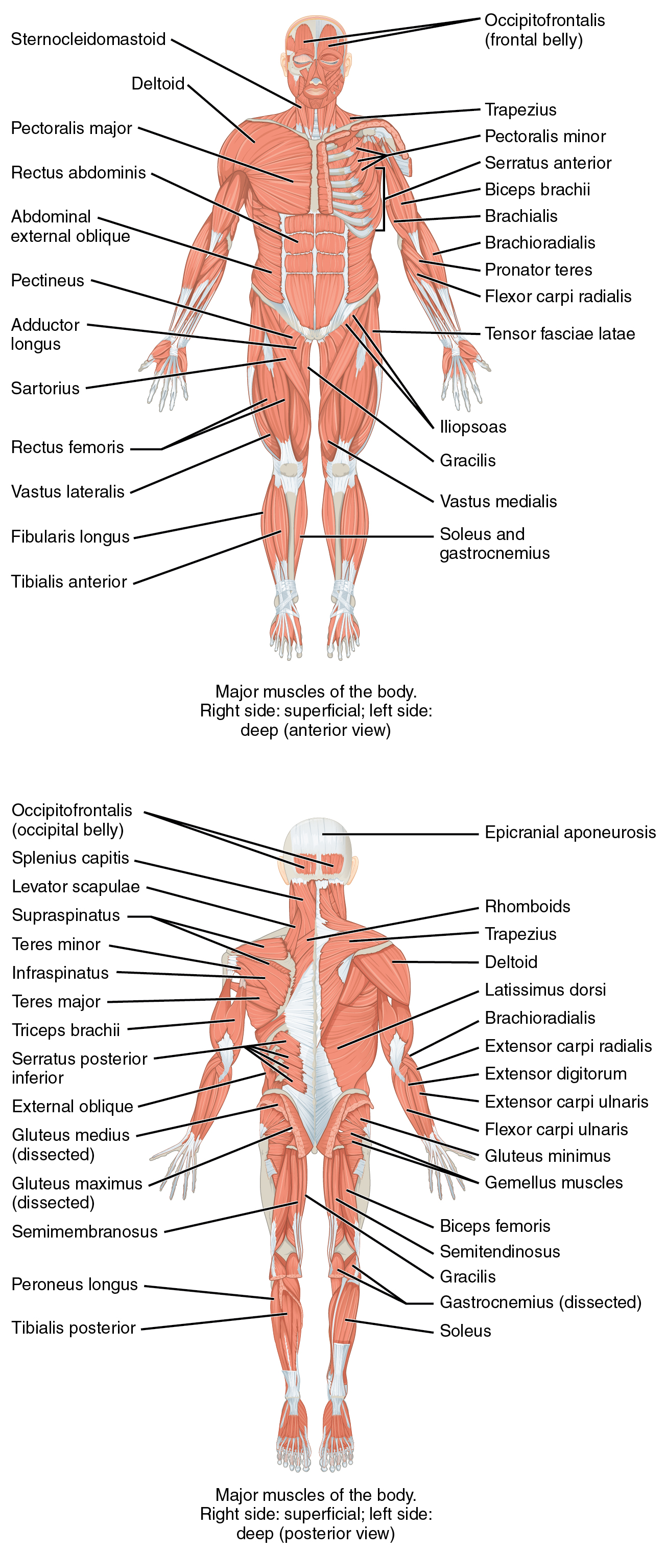
A complete list of muscles We've created muscle anatomy charts for every muscle containing region of the body: Upper limb Lower limb Head and neck Trunk wall Each chart groups the muscles of that region into its component groups, making your revision a million times easier.

Simple Human Muscles Diagram / Learn All Muscles With Quizzes And Labeled Diagrams Kenhub
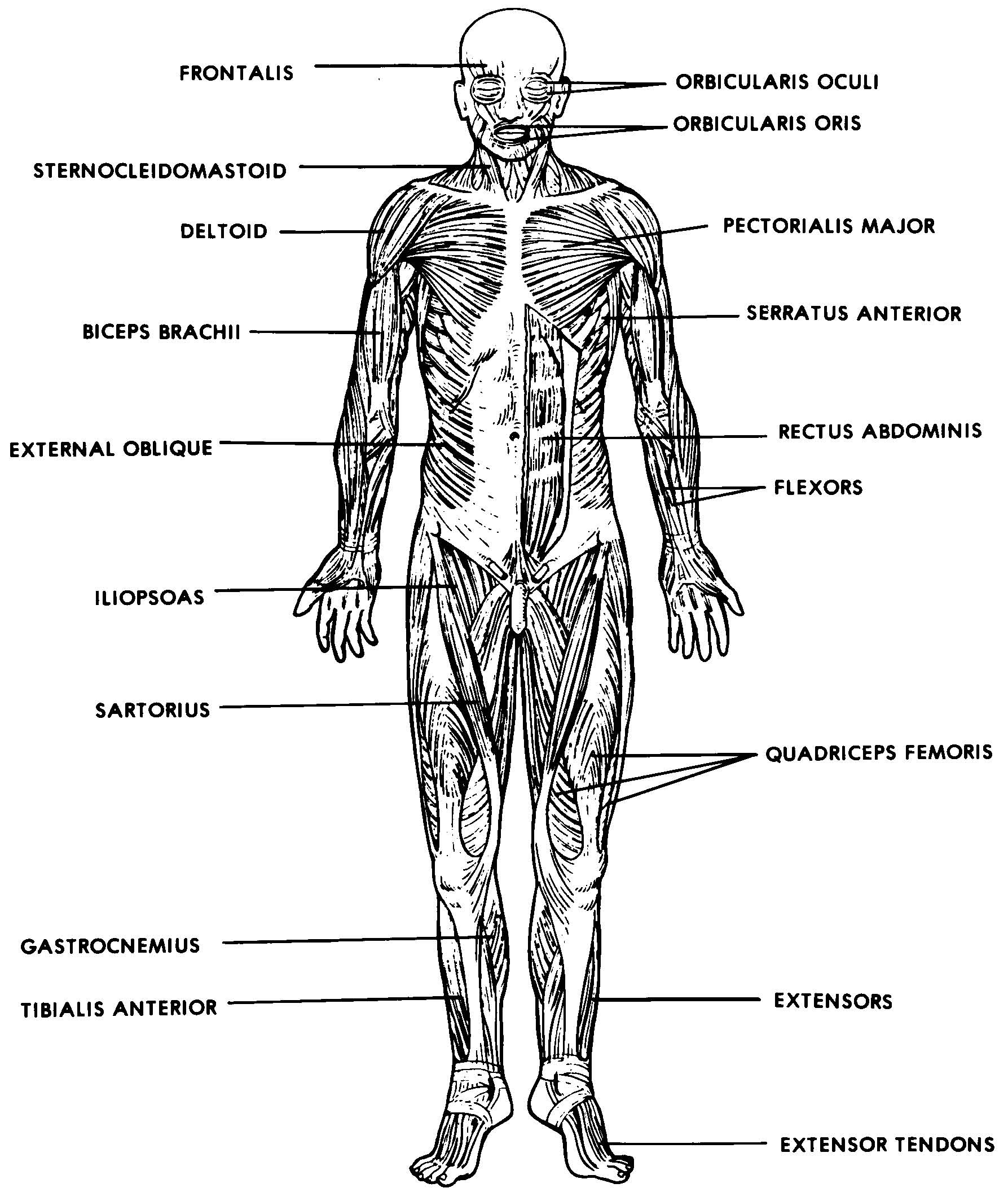
THE MUSCULAR SYSTEM COMPILED BY HOWIE BAUM 1 Muscles make up the bulk of the body and account for 1/3 of its weight.!! Blood vessels and nerves run to every muscle, helping control and regulate each muscle's function. The muscular system creates body heat and also moves the: Bones of the Skeletal system Food through Digestive system
4 human body muscles labeled Biological Science Picture Directory
The muscular system is made up of three types of muscle tissue: skeletal, cardiac and smooth. In this section, we'll focus on the skeletal muscles of the body involved in voluntary movement and maintaining posture. They are attached to bones via tendons and contract to cause movement at the joints. Learn more about the anatomy and functions of.
11.2 Naming Skeletal Muscles Anatomy and Physiology
The muscles in your upper and lower legs work together to help you move, support your body's weight and allow you to have good posture. They enable you to do big movements, like running and jumping. They also help you with small movements, like wiggling your toes. Leg muscle strains are common, especially in the hamstrings, quads and groin.

BodyPartChart Muscular System Front Labeled
Human Anatomy, 6/e. Kent Van De Graaff, Weber State University. Muscular System. Labeling Exercises. Muscles-Anterior View 1 Muscles-Anterior View 2 Muscles- Anterior View 3 Leg Muscles-Anterior View 1 Leg Muscles-Anterior View 2 Muscles-Posterior View 1
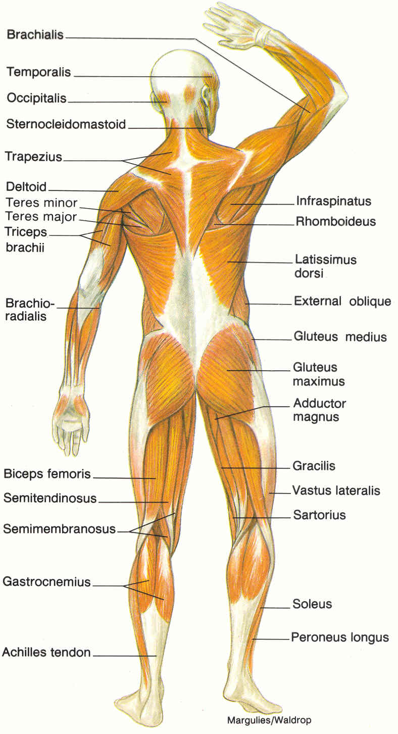
diagram of muscular system Biological Science Picture Directory
Muscle diagrams are a great way to get an overview of all of the muscles within a body region. Studying these is an ideal first step before moving onto the more advanced practices of muscle labeling and quizzes. If you're looking for a speedy way to learn muscle anatomy, look no further than our anatomy crash courses .