human cell diagram with labels
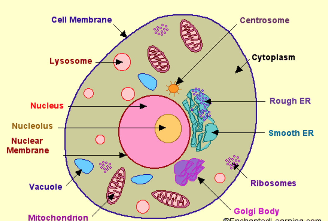
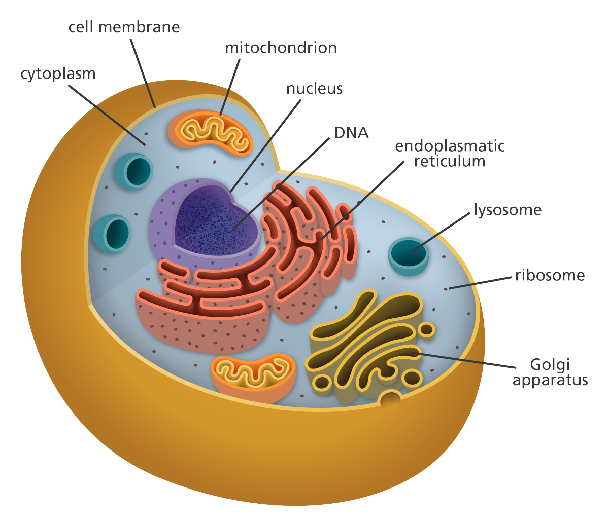
Diagram 1: The anatomical presentation of the human cell. Picture Source: www.printablediagram.com How many cells are in the human body ? Ans : Approx. 37.2 trillion cells What are the different parts of the human cells? How do these parts function? Cell membrane It is the outer covering of the cell, which consists of proteins and lipids.
Education Chart of Biology for Human Cell Diagram Best Acupuncture llc
The Human Cell Atlas is an international collaborative consortium that charts the cell types in the healthy body, across time from development to adulthood, and eventually to old age. This enormous undertaking, larger even than the Human Genome Project, will transform our understanding of the 37.2 trillion cells in the human body..
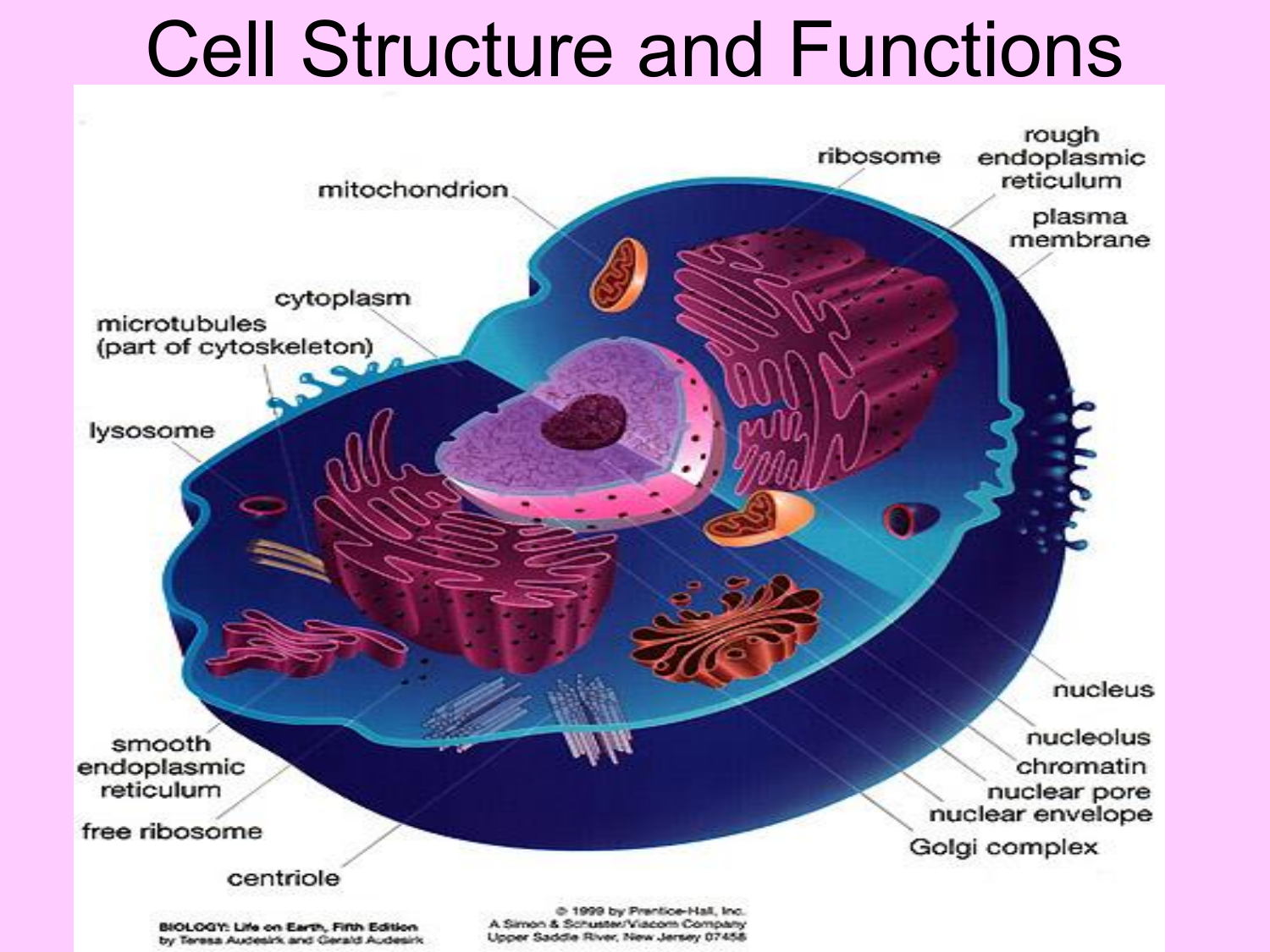
Cell Structure and Functions
Muscle tissue is made up of cells that have the unique ability to contract or become shorter. There are three major types of muscle tissue, as pictured in Figure 10.3.14 10.3. 14: skeletal, smooth, and cardiac muscle tissues. Skeletal muscles are striated, or striped in appearance, because of their internal structure.
Cell Structure and Function Part 1 The Organelles Medical Exam Prep
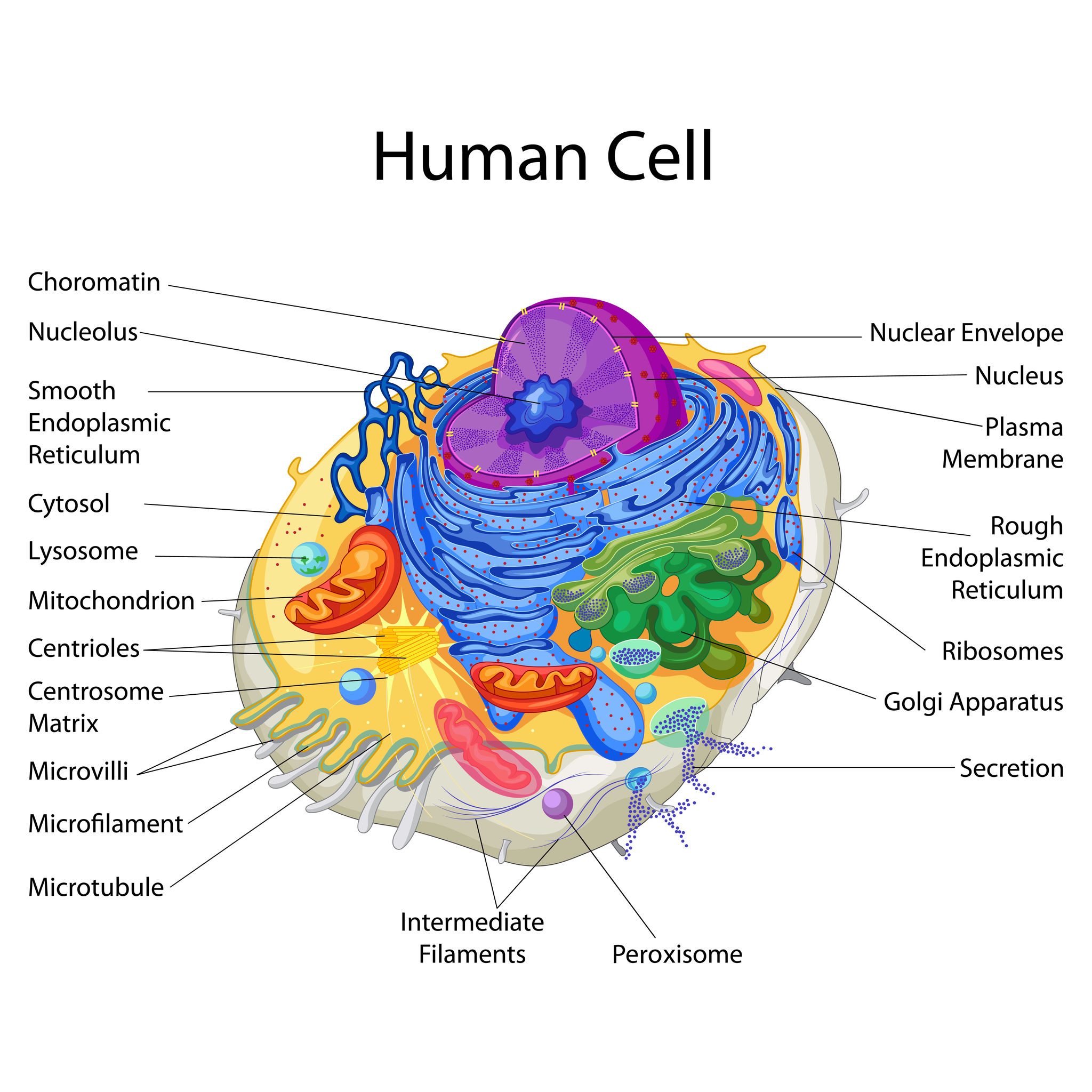
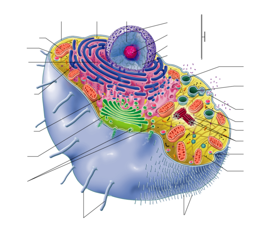
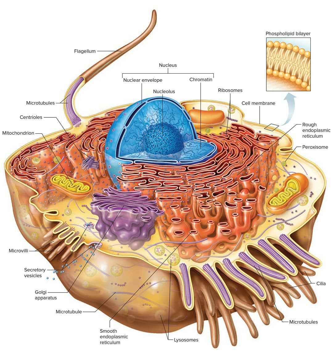
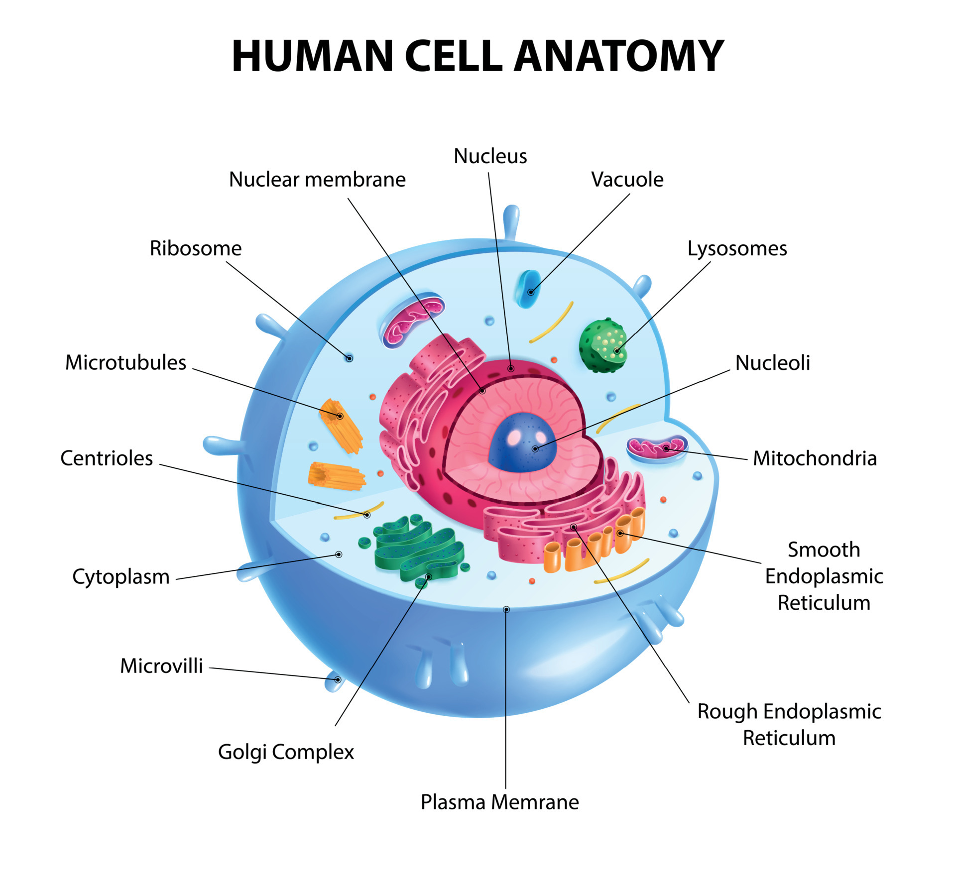
A human cell diagram provides a visual representation of the different components and organelles within a cell. An unlabeled human cell diagram, in particular, offers an excellent learning tool for students and researchers, encouraging them to identify and label the various parts independently.
Human cell diagram Etsy
Cell Structure Ideas about cell structure have changed considerably over the years. Early biologists saw cells as simple membranous sacs containing fluid and a few floating particles. Today's biologists know that cells are infinitely more complex than this. There are many different types, sizes, and shapes of cells in the body.
generalized human cell, labeled Diagram Quizlet
Interactive guide to stem cells and cell biology with 3D models and real microscopy data of GFP labeled hiPSCs.

Cell Nursing
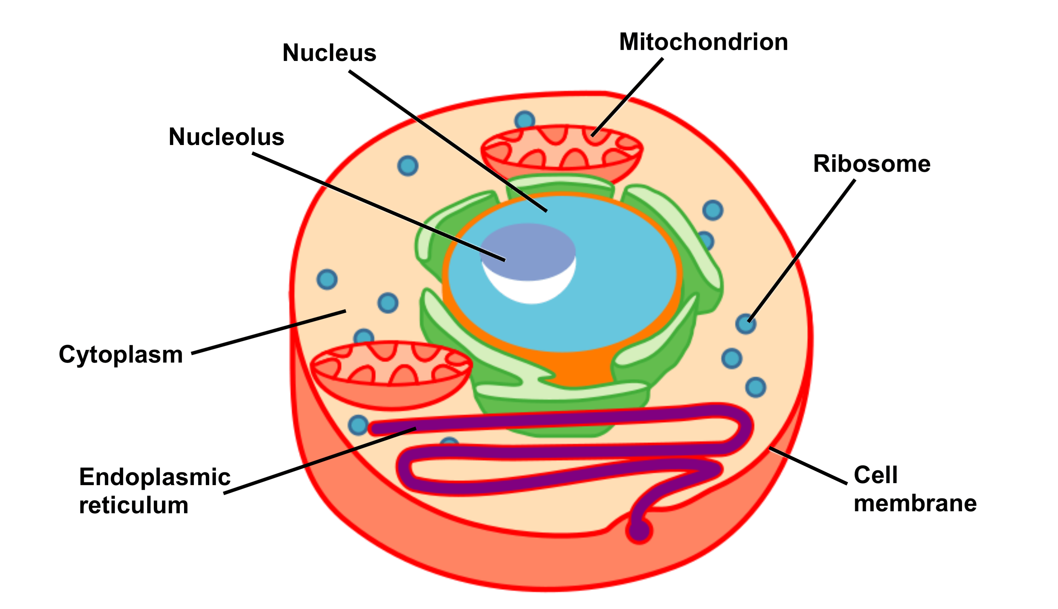
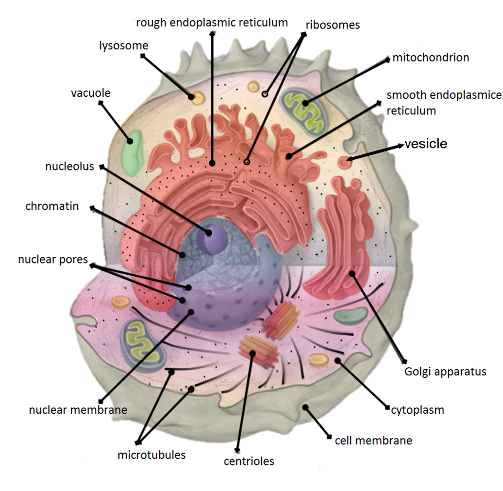
Key points: All cells have a cell membrane that separates the inside and the outside of the cell, and controls what goes in and comes out. The cell membrane surrounds a cell's cytoplasm, which is a jelly-like substance containing the cell's parts. Cells contain parts called organelles. Each organelle carries out a specific function in the cell.

human cell Diagram Quizlet
Cells of humans typically have a mass 400,000 times larger than the mass of a single mycoplasma bacterium, but even human cells are only about 20 μm across. It would require a sheet of about 10,000 human cells to cover the head of a pin, and each human organism is composed of more than 30,000,000,000,000 cells.
[DIAGRAM] Parts Of A Cell Diagram
The human (mammalian) cell is a complex structure able to carry out all the functions required to maintain cell life and also makes its contribution to homeostasis through the activities of the different organelles (small organs) within the cell. Figure 2.1 illustrates a generic cell filled with the liquid cytoplasm
Cell Biology, Cell Structure
The Glucose transporter 1 (GLUT1) is one of the most abundant proteins within the erythrocyte membrane and is required for glucose and dehydroascorbic acid (Vitamin C precursor) transport. It is widely recognized as a key protein for red cell structure, function, and metabolism. Previous reports highlighted the importance of GLUT1 activity within these uniquely glycolysis-dependent cells, in.
Human Cell Diagram 6406474 Vector Art at Vecteezy
A cell is the smallest living organism and the basic unit of life on earth. Together, trillions of cells make up the human body. Cells have three parts: the membrane, the nucleus, and the.

Human Cell Diagram, Parts, Pictures, Structure and Functions Diseases Pictures
The human cells typically contain 46 chromosomes (except mature sex cells which contain a haploid number of chromosomes, i.e., 23 chromosomes). The DNA molecules carry the master code for making all of the enzymes and other proteins of a cell. Thus they dictate both the structure and the function of the cells.
The Cell Theory & Structure HubPages
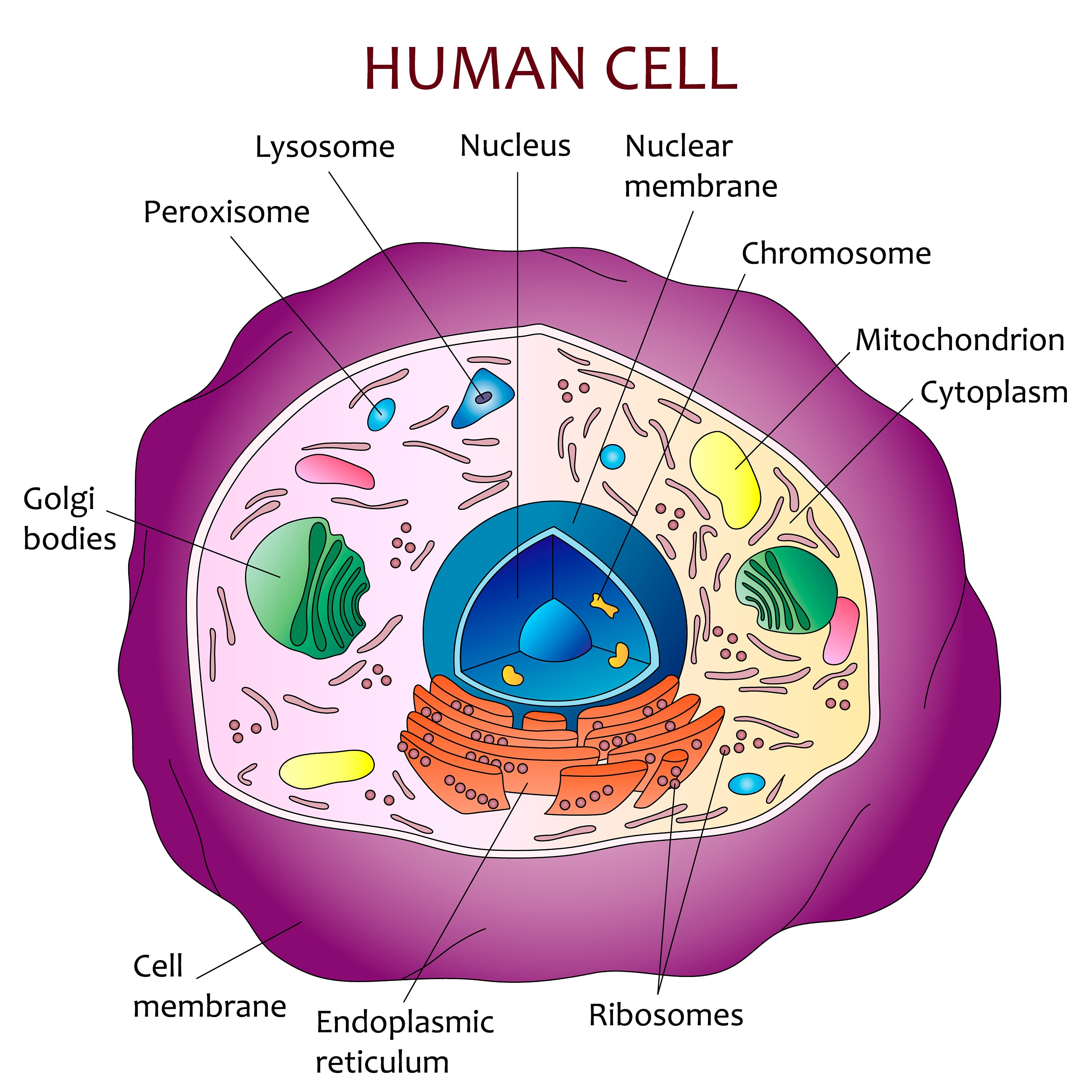
Diagram of the human cell illustrating the different parts of the cell. Cell Membrane The cell membrane is the outer coating of the cell and contains the cytoplasm, substances within it and the organelle. It is a double-layered membrane composed of proteins and lipids.
4.5 Cytoplasm and Cytoskeleton Human Biology
Human Cell Diagram, Parts, Pictures, Structure and Functions The cell is the basic functional in a human meaning that it is a self-contained and fully operational living entity. Humans are multicellular organisms with various different types of cells that work together to sustain life.

Labeled Diagram Human Cell
Division and differentiation in human cells When cells express specific genes that characterise a certain type of cell we say that a cell has become differentiated. Structure and replication of DNA

69,023 Human Cell Structure Images, Stock Photos & Vectors Shutterstock
Cell types Cells are broadly categorized into two types: eukaryotic cells, which possesses a nucleus, and prokaryotic cells, which lack a nucleus but still has a nucleoid region. Prokaryotes are single-celled organisms, whereas eukaryotes can be either single-celled or multicellular. [15] Prokaryotic cells Structure of a typical prokaryotic cell